What is the best UV?
- BEST OPTION: Convoy 8+ 365nm UV LED Flashlight with Patented Glass Filter
- BUDGET OPTION: Karrong Rechargeable 1200 Lumen 395nm UV Flashlight
- OPTION FOR INDOOR USAGE: Prime Upgraded Big Chip 396nm UV LED Light Lamp
What is woodwardfieser rule in UV spectroscopy?
These sets of rules to calculate the wavelength of maximum absorption or λmax of a compound in the ultraviolet-visible spectrum, based empirically have been called the Woodward-Fieser rules or Woodward’s-rules.
What are the applications of UV-visible spectroscopy?
Applications of UV-Vis spectroscopy
- DNA and RNA analysis. Rapidly determining the purity and amount of DNA and RNA is one of the most popular applications. ...
- Pharmaceutical analysis. The most popular applications of UV-Vis spectroscopy can be found within the pharmaceutical industry.
- Bacterial culture. ...
- Beverage analysis. ...
- Other applications. ...
What is the range of visible UV?
Ultraviolet-visible (UV-Vis) spectrophotometers use a light source to illuminate a sample with light across the UV to the visible wavelength range of the electromagnetic spectrum (typically 190 to 900 nm). The UV range normally extends from 100 to 400 nm, with the visible range from approximately 400 to 800 nm.
What does a UV-VIS spectrophotometer measure?
What does a UV-Vis spectrophotometer measure? UV-Vis and UV-Vis-NIR instruments measure the light absorbed, transmitted, or reflected by the sample across a certain wavelength range.
What is actually measured by UV-Vis absorption instrument?
A UV-Vis spectrophotometer measures the intensity of light transmitted through a sample compared to a reference measurement of the incident light source.
What information can be obtained from UV-Vis spectra?
Ultraviolet-visible (UV-Vis) spectroscopy is a widely used technique in many areas of science ranging from bacterial culturing, drug identification and nucleic acid purity checks and quantitation, to quality control in the beverage industry and chemical research.
Why is UV spectroscopy used?
UV–visible spectroscopy is routinely used in analytical chemistry for the quantitative determination of analytes, such as transition metal ions, highly conjugated organic compounds, and biological macromolecules. UV–visible is used to determine the size and concentration of NPs.
Why is UV spectroscopy used in pharmaceutical analysis?
UV spectrophotometers measure the visible regions of ultraviolet light and can provide valuable information, as well as detect any impurities, abou...
What are the applications of spectrophotometry?
In different fields, such as astronomy, molecular biology , chemistry and biochemistry, spectrophotometers are commonly used. Specification applica...
What is the range of UV spectroscopy?
UV-Vis is also considered a general procedure, since in the UV-visible wavelength spectrum, most molecules absorb light. The UV frequency is betwee...
Which lamp is used in UV spectroscopy?
Light with a wavelength range between 190 nm and 800 nm is radiated through the cuvette using a spectrometer and absorption spectrums are recorded....
What is the IR principle?
The principle of IR spectroscopy utilises the idea that molecules appear to absorb unique light frequencies that are typical of the molecules’ corr...
What is UV VIS spectroscopy and how does it work?
UV-Vis is a quick , convenient, and inexpensive way of determining the solution concentration of an analyte. In UV-Vis, a beam travels through a so...
What is UV visible spectroscopy?
Ultraviolet (UV)-visible spectroscopy is a type of absorption spectroscopy in which UV-visible light is absorbed by the molecule. Absorption of the UV-visible radiations results in the excitation of the electrons from lower to higher energy levels. In organic molecules only certain functional groups (chromophores) that contain valence electrons of low excitation energy can absorb ultraviolet and visible radiation. C-Cyts represent an ideal target molecule for UV-visible spectroscopy because of the large absorption of heme groups. The strong UV-visible absorption bands of the heme originate from the π→π* transitions, providing information about the type of heme, the oxidation, and the spin state of the central iron ion. UV-visible spectroscopy allows in vivo measurements of biofilms under physiologically relevant conditions (Fig. 4D ). In order to detect all the cytochromes (OMCs and inner membrane cytochromes) along the biofilm thickness without any spatial distinction growing the EABs on a transparent electrode (indium tin oxide) is suggested. 78 Moreover, by combining different experimental set-ups is possible to obtain a UV-visible spectrum of the OMCs only confined in the proximity of the electrode surface.
What is UV VIS?
UV–vis is a commonly used technique to characterize nanoparticles. This technique allows to confirm the nanoparticles formation by measuring the Surface Plasmon Resonance (SPR). This procedure can provide information about the size, stability, and aggregation of the NPs [4].
How to measure analyte interactions with MIPs?
UV/Vis spectroscopyis one of the most simplified and economical methods for examining analyte interactions with MIPs where only the change in absorbance is measured as a function of wavelength. The technique is versatile and gives rapid response regarding quantitative information on template binding. Besides pure sensing application, this method is quitesuitable for screening [36]MIPs and choosing the finest polymer composition. With the help of the UV/Vis spectrum, the thorough mechanism of complexation between templates, monomer, and cross-linker during polymerization can also be better understood. It has been observed that after complexation, an absorbance shift toward shorter wavelengths takes place. The procedure makes it easy to compare the spectrum of free template and functional monomer with that of the complex formed. This strategy is equally suitable for monitoring metal polymer complexation in visible regions [37]. Although UV/Vis spectroscopy is not as selective as the fluorescence method, it is nevertheless quite suitable for designing low-cost MIP sensors with moderate sensitivity.
What is FUV spectroscopy used for?
Moreover, FUV spectroscopy can be utilized for qualitative and quantitative analyses of various liquid and solid samples, because each molecule shows a characteristic FUV spectrum with strong absorption, and intensities and wavelengths of FUV bands are very sensitive to changes in concentration, temperature, pH, and so on [ 46–50].
What are the advantages of FUV spectroscopy?
The most fundamental advantage of FUV spectroscopy is that it contains unique information about the electronic transitions and structure of molecules. One can obtain knowledge about them that is not accessible by any other spectroscopy.
Why are C-cyts used in UV spectroscopy?
C-Cyts represent an ideal target molecule for UV-visible spectroscopy because of the large absorption of heme groups. The strong UV-visible absorption bands of the heme originate from the π→π* transitions, providing information about the type of heme, the oxidation, and the spin state of the central iron ion.
How is light absorbed by a sample measured?
The physical principles underlying this method are straightforward, making the instrumentation simple and robust. Light of known wavelength and intensity is directed at the sample and its final intensity, after passing through, is measured by a detector. By comparing the incident radiation (I0) and the transmitted radiation (I), the amount of light absorbed by the sample at that particular wavelength can be easily calculated. Using the Beer–Lambert law, this absorption can be used to measure concentrations of known solutes:
What is UV spectroscopy?
UV Vis spectroscopy is a type of absorption spectroscopy in which a sample is illuminated with electromagnetic rays of various wavelengths in the ultraviolet (UV) and visible (Vis) ranges. Depending on the substance, the UV or visible light rays are partially absorbed by the sample. The remaining light, i.e. the transmitted light, is recorded as a function of wavelength by a suitable detector. The detector then produces the sample's unique UV Vis spectrum (also known as the absorption spectrum).
How to analyze a compound with UV spectroscopy?
Molecules can be analyzed using UV Vis spectroscopy if they possess any functional group or conjugation, or if they produce a color complex. As inorganic compounds do not contain any functional group or conjugation, the common method for analyzing them is by reaction with a suitable compound. This produces a color complex whose absorbance can be photometrically measured in the visible region and correlated with its actual concentration. For example, iron is commonly analyzed by a reaction with 1, 10-phenthroline to produce a red color complex. The absorbance of the complex is measured at 570 nm to estimate iron concentration.
How to measure transmittance in a spectrophotometer?
In a spectrophotometer the transmittance is measured by dividing the intensity spectrum of light transmitted through a sample (I) by the intensity spectrum of light transmitted through the blank (I 0 ).
Why is the sample compartment open in UV spectrophotometers?
The sample compartment in UV Vis array spectrophotometers is open due to the fact that array instruments use reverse optics and the simultaneous detection of all wavelengths of the spectrum.
What happens to the absorption of UV light?
The absorption of UV light results in electronic transitions from lower energy levels to higher energy levels. Absorption of ultraviolet radiation in organic molecules is restricted to certain functional groups (chromophores) that contain valence electrons of low excitation energy. The molecular transitions/interactions that take place due to UV absorption are:
What are the different types of spectroscopic techniques?
The spectroscopic techniques commonly used for chemical analysis are atomic spectroscopy, ultraviolet and visible spectroscopy (UV Vis spectroscopy), infrared spectroscopy, Raman spectroscopy and nuclear magnetic resonance .
With UV Vis Spectrophotometry
Color measurement is an important parameter in the quality control of any manufactured goods. The determination of color was in the hands of specially trained experts for a long time. However, this has since been replaced by a technologically more advanced solution such as UV Vis spectrophotometry.
Contents
1 Introduction to Color 1.1 Colors of the light 1.2 Transmittance, Absorbance and Reflectance 2 How we See the Light 3 Color Numbers 3.1 Yellowness Index 3.2 APHA, Pt/Co, Hazen 3.3 Saybolt 3.4 Gardner 3.5 EBC 3.6 ASBC 3.7 Hess-Ivess 4 Conclusion 5 Mettler-Toledo Color Measurements 5.1 Stand-Alone methods 5.2 LabX Methods 6 Tips and Tricks
What is the wavelength of a UV spectrophotometer?
Ultraviolet visible (UV-Vis) spectrophotometers use a light source to illuminate a sample with light across the UV to the visible wavelength range (typically 190 to 900 nm). The instruments then measure the light absorbed, transmitted, or reflected by the sample at each wavelength. Some spectrophotometers have an extended wavelength range, into the near-infrared (NIR) (800 to 3200 nm).
What light source is used for UV spectroscopy?
Unfortunately, such a source does not exist. Two different light sources have historically been used in UV-visible spectrophotometers: – The deuterium arc lamp was used to provide a good intensity continuum in the UV region and useful intensity in the visible region – The tungsten-halogen lamp yielded good intensity over the entire visible range and part of the UV spectrum More recently, a single Xenon flash lamp has been used more widely. The use of a Xenon flash lamp as a single source has significant advantages over the use of the two conventional lamps. Deuterium (D
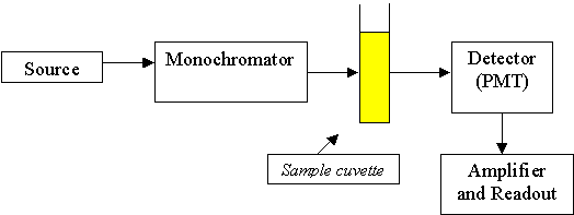
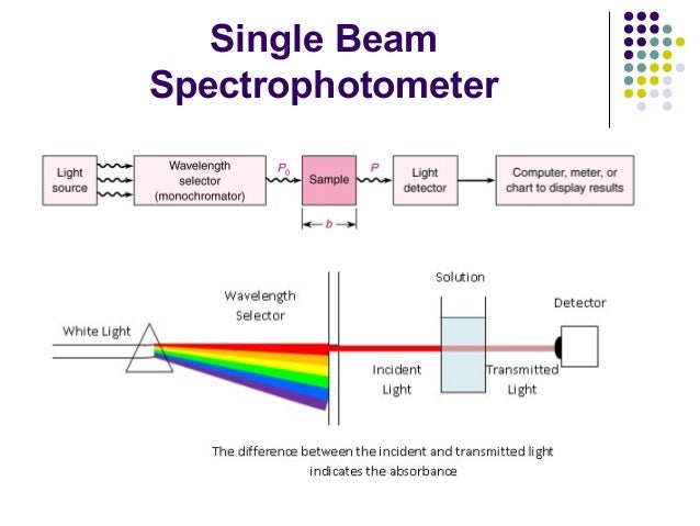
What is a single monochromator spectrophotometer?
A single monochromator spectrophotometer is used for general-purpose spectroscopy and can be integrated into a compact optical system. Figure 13 shows a schematic diagram of a single monochromator optical system. A single monochromator spectrophotometer cannot select the wavelengths of light as narrowly as a double monochromator system, but this ability may not be required for many applications, for example when measuring samples that have broad absorption peaks.
What wavelength of light is used to transfer electrons from the oxygen atom to the C-O bond?
Figure 2. Electronic transitions in formaldehyde. UV light at 187 nm causes excitation of an electron in the C-O bond and light at 285 nm wavelength causes excitation and transfer of an electron from the oxygen atom to the C-O bond.
How to determine the wavelength of electromagnetic radiation?
Because radiation acts as a wave, it can be classified in terms of either waveleng th or frequency, which are related by the following equation: ν = c/λ where ν is frequency (in seconds), c is the speed of light (3 × 10⁸ms-1), and λ is wavelength (in meters). In UV-Vis spectroscopy, wavelength is usually expressed in nanometers (1 nm = 10-9m). It follows from the equations that radiation with shorter wavelength has higher energy, and, for UV-Vis spectroscopy, the low (short)
When light passes through or is reflected from a sample, the amount of light absorbed is the difference between the?
When light passes through or is reflected from a sample, the amount of light absorbed is the difference between the incident radiation (I
How does a monochromator work?
To narrow the light down to a selected wavelength band, the light is passed through a monochromator. A monochromator consists of: – An entrance slit, – A dispersion device, to spread the light into different wavelengths (like a rainbow) and allow the selection of a nominated band of wavelengths, and – An exit slit where the light of the nominated wavelengths passes through and onto the sample. An easy way to think about a monochromator is to think of a room, with the sun shining through a window. The sunlight hits a prism that separates the white light into a rainbow. The rainbow falls onto a window on the opposite side of the room. As the prism is turned, light of different colors i.e. different wavelengths, pass out of the room through the window. Ideally, the output from a monochromator is light of a single wavelength. In practice, however, the output is always a band of wavelengths. Most spectrophotometers on the market today contain holographic gratings as the dispersion device. These optical components are made from glass, onto which extremely narrow grooves are precisely etched onto the surface. The dimensions of the grooves are of the same order as the wavelength of light to be dispersed. Finally, an aluminum coating is applied to create a reflective surface. Interference and diffraction of the light falling on the grating is reflected at different angles, depending on the wavelength. Holographic gratings yield a linear angular dispersion with wavelength and are temperature insensitive. However, they reflect light in different orders, which overlap (see Figure 12). As a result, filters must be used to ensure that only the light from the desired reflection order reaches the detector. A concave grating disperses and focuses light simultaneously.
What is UV/VIS spectroscopy?
Ultraviolet and visible light range (UV/VIS) is widely applied in research, production and quality control for the classification and study of substances. UV/VIS spectroscopy is based on the absorption of light by a sample. Depending on the amount of light and its wavelength absorbed by the sample, valuable information can be obtained, such as the purity of the sample. Moreover, the amount of absorbed light is related to the amount of sample, and thus, quantitative analysis is possible by optical spectroscopy. This article more specifically explores techniques when using a spectrophotometer to determine concentration of an analyte. A UV/VIS spectrophotometer measures the intensity of light passing through a sample solution in a cuvette, and compares it to the intensity of the light before it passes through the sample. The main components of a UV/VIS spectrophotometer are a light source, a sample holder, a dispersive device to separate the different wavelengths of the light and a suitable detector. This instrument measures Transmittance which is the ratio of the transmitted intensity I to the original intensity of light. An important derived (calculated) variable also reported by the instrument is the Absorbance which is defined as A = −log (Transmittance).
When using a spectrophotometer to determine concentration of a sample solution of unknown concentration by UV/VIS?
When using a spectrophotometer to determine concentration of a sample solution of unknown concentration by UV/VIS spectroscopy, a calibration line must first be created . This is done by measuring the light absorption of several standard solutions of different , known concentrations at a predefined, fixed wavelength. The below calibration line is obtained:
How do scanning spectrophotometers measure transmittance?
The grating is rotated in order to individually select each wavelength that is then sent through a cuvette. The transmittance at this specific wavelength is recorded. The whole spectrum is obtained by continuously changing the wavelength of light (i.e. scanning) incoming onto the sample solution by rotating the grating. Alternately, in Array Spectrophotometers, the sample is illuminated by a light beam consisting of all spectral components of the UV/ VIS range. The sample in the cuvette absorbs all wavelengths simultaneously and the transmitted light is diffracted and then detected by a CCD sensor. Measuring the whole UV/VIS spectrum is generally faster than using a conventional scanning spectrophotometer since the spectrum is recorded simultaneously at all wavelengths. Moreover, an array detector has an integrating function which accumulates individual measurements to enhance the signal, leading to a strongly increased signal to noise ratio, and thus to an improved signal quality of the measured spectrum. Array Spectrophotometers present an innovative approach to speed up full spectrum scan based on reverse optics technology. The robust design without any moving optical parts ensures very good optical performance.
What is the wavelength of a Xenon flash lamp?
The light source consists of a Xenon flash lamp for the ultraviolet (UV) as well as for the visible (VIS) and near-infrared wavelength regions covering a spectral range from 190 up to 1100 nm. The lamp flashes are focused on a glass fiber which drives the beam of light onto a cuvette containing the sample solution.
Is UV absorbance a function of wavelength?
Absorbance as a function of wavelength. In general, a UV/VIS spectrum is graphically represented as absorbance as a function of wavelength. The advantage of this representation is obvious; the height of the absorption peaks is directly proportional to the concentration of the species. The calculation of concentration is governed by ...

How Does A Uv-Vis Spectrophotometer Work?
Uv-Vis Spectroscopy Analysis, Absorption Spectrum and Absorbance Units
- UV-Vis spectroscopy information may be presented as a graph of absorbance, optical density or transmittance as a function of wavelength. However, the information is more often presented as a graph of absorbance on the vertical y axis and wavelength on the horizontal xaxis. This graph is typically referred to as an absorption spectrum; an example is...
Strengths and Limitations of Uv-Vis Spectroscopy
- No single technique is perfect and UV‑Vis spectroscopy is no exception. The technique does, however, have a few main strengths listed below that make it popular. 1. The technique isnon‑destructive, allowing the sample to be reused or proceed to further processing or analyses. 2. Measurements can be made quickly, allowing easy integration into experimental protocols. 3. …